
Liene Znotina
Femur (posterior view) (English)
Femur (posterior view) (English)
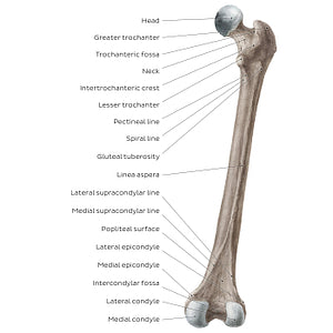
The lesser trochanter is clearly seen on the posterior view, separated from the greater trochanter by the intertrochanteric crest. The intertrochanteric crest and intertrochanteric line (anterior view) mark the transition between the neck of the femur and the shaft of the femur. Found on the medial surface of the greater trochanter is the crescent-shaped depression known as the trochanteric fossa. Seen below the lesser trochanter are the 3 small bony ridges called pectineal line, spiral line and gluteal tuberosity. These 3 ridges converge inferiorly to form the linea aspera that runs along the entire shaft of the femur. Near the distal end of the femur, the linea aspera diverges into 2 ridges: the medial and lateral supracondylar lines. The posterior surface of the distal end of the femur provides a better visual of the medial and lateral condyles, which in this view are separated by the intercondylar fossa.
Precio habitual
$7.56 USD
Precio habitual
Precio de oferta
$7.56 USD
Precio unitario
por
No se pudo cargar la disponibilidad de retiro

#8F806D
#57473C y #C7BCB3