
Liene Znotina
Femur (anterior view) (English)
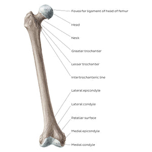
Femur (anterior view) (English)
The anterior view of the femur features several important landmarks. The most proximal portion of the head of the femur features a small dimple, known as the fovea for the ligament of head of femur. Below the neck of the femur are the greater and lesser trochanters, with the intertrochanteric line spanning between them. These bony prominences act as important attachment sites for the muscles of the hip and thigh. The distal end of the femur contains the medial and lateral condyles that articulate with the tibia, as well as the medial and lateral epicondyles above them. Between the medial and lateral condyle is the patellar surface of the femur, which as its name suggests, articulates with the patella, contributing to the formation of the knee joint. Notice how some of these landmarks are better seen on the posterior view (2nd image), such as the lesser trochanter, and the medial and lateral condyles.
Precio habitual
$7.56 USD
Precio habitual
Precio de oferta
$7.56 USD
Precio unitario
por
No se pudo cargar la disponibilidad de retiro

#8C7D72
#483A32 y #C8BCB3