Samantha Zimmerman
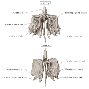
Ethmoid bone (anterior and posterior views) (Latin)
Ethmoid bone (anterior and posterior views) (Latin)
The os ethmoidale is an unpaired bone situated in the midline of the cranium. It consists of a centrally positioned crista galli, which is continued inferiorly by the lamina perpendicularis.On each side of the midline there is a spongy lateral mass, often referred to as the labyrinthus ethmoidalis. Each mass consists of numerous air-filled cellulae ethmoideae. Thus, each mass is known as the sinus ethmoidei. The lateral wall of the sinus presents a saccular extension called the bulla ethmoidalis, the largest of cellulae ethmoideae. Ethmoidal masses feature a hook-like projection pointing inferiorly called the processus uncinatus. This process forms a part of the wall of the sinus maxillaris and is often confused as a part of the maxilla rather than of os ethmoidale. The lateral surface of ethmoidal masses faces the orbita, forming a part of its medial wall. The medial surface of each mass faces the lamina perpendicularis. From an anterior view, it is visible how a separate bony lamina called the concha media nasi extends inferiorly from the root of each mass. The posterior view allows the appreciation of the concha superior nasi as well. The conchae project into the cavitas nasi increasing its surface. Between the conchae is the meatus medius nasi, which drains the cellulae ethmoidales into the cavitas nasi.
Precio habitual
$7.56 USD
Precio habitual
Precio de oferta
$7.56 USD
Precio unitario
por
No se pudo cargar la disponibilidad de retiro


#897C73
#4E4138 y #C5BCB4

