Paul Kim
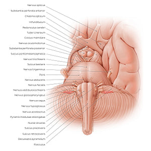
Anterior view of the brainstem (Latin)
Anterior view of the brainstem (Latin)
Along its ventral surface, the mesencephalon is characterized by two prominences known as the pedunculi cerebri which connect the cerebral hemispheres (hemispheria cerebri) to the brainstem (truncus encephali). Between each pedunculus cerebri is a shallow depression, the fossa interpeduncularis; the substantia perforata posterior forms the floor of this fossa, while its contents include the nn. oculomotorii (CN III) and corpora mamillaria. The pons is limited superiorly by the sulcus pontomesencephalicus (sulcus pontinus superior) and ends inferiorly at the sulcus bulbopontinus (sulcus pontinus inferior). When viewed from the ventral aspect, the pons resembles a dome-like structure with numerous horizontal striations across its surface. A shallow depression runs along the vertical axis known as the sulcus basilaris, which houses the a. basilaris. The n. trigeminus (CN V) emerges from the lateral aspect of the pons while the n. abducens (CN VI), n. facialis (VII) and n. vestibulocochlearis (CN VIII) arise from the sulcus bulbopontinus. The fissura mediana anterior divides the ventral medulla oblongata into symmetrical halves and is bordered on either side by the pyramides medullares. Lateral to each pyramis is another prominent bulge, the olive (oliva), which corresponds to the location of the nuclei olivares. At its distal end, the fissura mediana anterior is interrupted by criss-crossing fibres known as the decussatio pyramidum, which marks the termination of the medulla oblongata. The lower four cranial nerves (CN IX-XII) emerge from the anterior surface of the medulla oblongata.
Precio habitual
$7.56 USD
Precio habitual
Precio de oferta
$7.56 USD
Precio unitario
por
No se pudo cargar la disponibilidad de retiro


#E16D5F
#976557
#943625
#734F47
#F1948B y #D2B3AA

