
Paul Kim
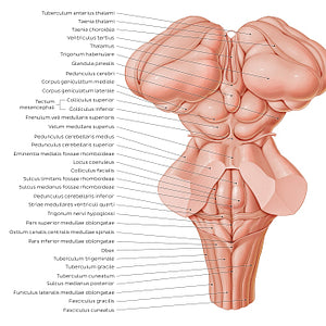
Surface anatomy of the brainstem (Latin)
Surface anatomy of the brainstem (Latin)
The posterior part (tectum) of the midbrain (mesencephalon) has two raised, round protrusions that are collectively known as the lamina quadrigemina. This complex consists of the colliculi superioris et inferioris. Each colliculus inferioris is separated from the contralateral counterpart by the frenulum veli medullaris superioris. The dorsal aspect of the pons and upper part of medulla oblongata forms the floor of the ventriculus quartus, forming a large landmark known as the fossa rhomboidea. The upper fossa rhomboidea is divided in half by a sulcus medianus, with each half being further divided again by a parallel line, the sulcus limitans. Between the sulcus medianus and sulcus limitans is the eminentia medialis, while the region lateral to the sulcus limitans is the upper portion of the area vestibularis. The ncl. caeruleus/locus caeruleus is also found on the upper fossa rhomboidea. The cranial/superior limit of the pons is formed by a structure which contributes to the formation of the tegmen ventriculi quarti, the velum medullare superius, while the inferior border is formed by the striae medullares ventriculi quarti. The dorsal/posterior surface of the medulla oblongata is divided into an open/pars superior, which contains the caudal half of the ventriculus quartus, and a closed/pars inferior, which contains the canalis centralis that continues into the medulla spinalis. Structures of the lower fossa rhomboidea within the superior part of the medulla oblongata include the trigonum nervi hypoglossi and trigonum nervi vagi, obex and lower portion of the area vestibularis. The dorsal aspect of the pars inferior medullae oblongatae is marked by a sulcus medianus posterior, which is bounded on either side by a raised structure known as the tuberculum gracile which is in turn bordered laterally by the tuberculum cuneatum. These landmarks are continuations of the fasciculus gracilis/cuneatus of the medulla spinalis.
Preço normal
$7.56 USD
Preço normal
Preço promocional
$7.56 USD
Preço unitário
por
Não foi possível carregar a disponibilidade de retirada.

#C4623B
#94655A
#692720
#736F6D
#F2AB97 e #D1B3A9