Paul Kim
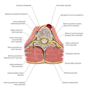
Spinal cord in situ (Latin)
Spinal cord in situ (Latin)
On exiting the foramen intervertebrale, each n. spinalis divides into a r. anterior and r. posterior. The r. posterior extends in a posterior direction and further divides into a r. medialis/lateralis which provide innervation to the mm. dorsi produndi (mm. epaxiales) as well as an associated narrow strip of overlying skin. The r. anterior provides motor innervation (somatic or visceral) to the rest of the body related to that segmental level, often intermingling with other spinal nerves leading to the formation of the major somatic plexuses (plexus cervicalis, brachialis, lumbalis and sacralis). Just before the bifurcation into rr. anterior and posterior, the n. spinalis also gives rise to a r. meningeus/recurrens which re-enters the foramen intervertebrale to supply the meninges and other structures of the canalis vertebralis. Communicating with the r. anterior of the nn. spinales are the rr. communicantes grisei/albi which convey sympathetic nerve fibers. The rr. anteriores of nn. spinales T1-L2 give off the r. communicans albus which carries preganglionic sympathetic fibers from the cornu lateralis of the spinal cord to an adjacent ganglion sympatheticum. Here they can either synapse with a postganglionic sympathetic neuron at the same or a superior/inferior level via the truncus sympatheticus, or pass through the ggl. sympatheticus (without synapsing) continuing as a preganglionic nerve fiber; in all cases nerve fibers of the truncus sympatheticus re-enter the nn. spinales via a r. communicans griseus.
Normaler Preis
$7.56 USD
Normaler Preis
Verkaufspreis
$7.56 USD
Grundpreis
pro
Verfügbarkeit für Abholungen konnte nicht geladen werden


#BC363E
#5A8DA1
#A0202B
#5F4B38
#FDEC75 und #CBB5AE

