Paul Kim
Spinal cord: Cross section (Gray matter) (Latin)
Spinal cord: Cross section (Gray matter) (Latin)
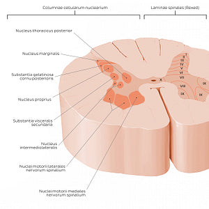
Neurons of the cornu posterius can be subdivided into four groups (nuclei): the ncl. marginalis (Rexed lamina I), substantia gelatinosa (Rexed lamina II), ncl. proprius (Rexed laminae III, IV, V), and ncl. thoracicus posterior (Rexed lamina VII). These nuclei function to receive and process sensory information carried by the radix posterior nervi spinalis. The cornu lateralis contains the ncll. intermediolateralis (Rexed lamina VII), which is a group of preganglionic sympathetic neurons. The ncl. intermediolateralis is present only at the level of T1-L3 segments and functions to provide sympathetic innervation, which is transported to the truncus sympathicus via the rami communicantes albus. Between S2-S4 is a ncl. parasympathicus sacralis which is also considered to belong to Rexed lamina VII.The neurons of the cornu anterius are responsible for motor innervation, carried by the radix anterior nervi spinalis. These neurons are divided into three groups: ncl. mediales (Rexed lamina IX), ncl. centrales (Rexed lamina IX) and ncl. laterales (Rexed lamina IX). The ncl. medialis extends almost entirely throughout the length of the medulla spinalis while the ncl. centralis and ncl. lateralis are only present in some segments. The ncl. centralis is found at the partes cervicalis et lumbosacralis medullae spinalis and the ncl. lateralis is present only in the regions of the medulla spinalis responsible for motor innervation to the upper and lower limbs (C4 to T1 and L2 to S3). Rexed lamina X is located around the canalis centralis at the commissura grisea and is associated with the systema nervosum autonomicum.
Normaler Preis
$7.56 USD
Normaler Preis
Verkaufspreis
$7.56 USD
Grundpreis
pro
Verfügbarkeit für Abholungen konnte nicht geladen werden


#CB6034
#9C715F
#8B3612
#6E5950
#F6936F und #CCB4AA

