Samantha Zimmerman
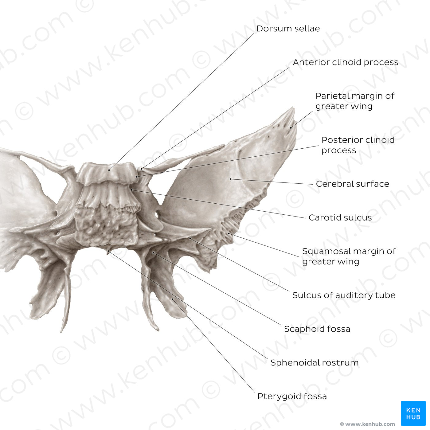
Sphenoid bone (posterior view) (English)
Sphenoid bone (posterior view) (English)
The posterior surface of the sphenoid body is rough, featuring a prominent landmark: dorsum sellae. The dorsum sellae articulates with the occipital bone and forms the clivus. The inferolateral angles of the dorsum present small posterior clinoid processes, which forms one of the attachment points for the tentorium cerebelli. In the midline, the body presents a triangular process called the sphenoidal rostrum which points towards the ala of vomer. The posterior view allows a better appreciation of the root of the lesser wing. This bony process spirals from the sphenoid body, forming the anterior clinoid process, after which it continues laterally and fuses with the greater wing. The irregular appearance of the root of the greater wing is also noticeable from this perspective. This area features the sulcus of auditory tube which houses the cartilaginous auditory tube. The cerebral surface of greater wing contributes to the middle cranial fossa, lodging a part of the temporal lobe of the brain. The ridged parietal and squamosal margins of the greater wings are visible, which serve for the articulations with the parietal and temporal bones respectively.From this aspect, the plates of the pterygoid process present as concave. At their origin there is a shallow scaphoid fossa which serves as the attachment site for the tensor veli palatini muscle. The very concavity of the lateral plate comprises an obtuse-angled pterygoid fossa which provides the attachment site for the medial pterygoid muscle.
Normaler Preis
$7.56 USD
Normaler Preis
Verkaufspreis
$7.56 USD
Grundpreis
pro
Verfügbarkeit für Abholungen konnte nicht geladen werden


#897C73
#4D4137 und #C5BCB4

