Samantha Zimmerman
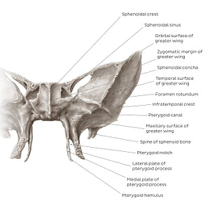
Sphenoid bone (anterior view) (English)
Sphenoid bone (anterior view) (English)
The anterior surface of the sphenoid body features the sphenoidal crest in the midline via which it articulates with the perpendicular plate of the ethmoid bone, contributing to the formation of the nasal septum. On each side of the crest is the sphenoidal concha which partially encloses the sphenoidal sinus. The superolateral part of the sinus remains open and communicates with the nasal cavity.The root of the greater wing features two openings: the foramen rotundum and pterygoid canal. The foramen rotundum is traversed by the maxillary branch of trigeminal nerve (CN V2), while the pterygoid (Vidian) canal transmits the artery and nerve of pterygoid canal. From an anterior perspective the orbital, temporal and maxillary surfaces of the greater wing are visible. The orbital surface contributes to the lateral part of the orbit. The temporal surface faces laterally and is divided by the infratemporal crest into superior and inferior portions. The superior portion contributes to the wall of the temporal fossa, while the inferior portion forms a part of the infratemporal fossa. The maxillary surface faces the maxilla.The spine of sphenoid bone points inferiorly from the lower margin of the temporal surface, providing the attachment site for the sphenomandibular ligament.The pterygoid process extends inferiorly from the root of the greater wing. It bifurcates into the lateral and medial plates, between which is a space called the pterygoid notch. The very tip of the medial plate is called the pterygoid hamulus.
Normaler Preis
$7.56 USD
Normaler Preis
Verkaufspreis
$7.56 USD
Grundpreis
pro
Verfügbarkeit für Abholungen konnte nicht geladen werden


#887C73
#4E4136 und #C4BCB4

