
Paul Kim
Knee joint - sagittal (English)
Knee joint - sagittal (English)
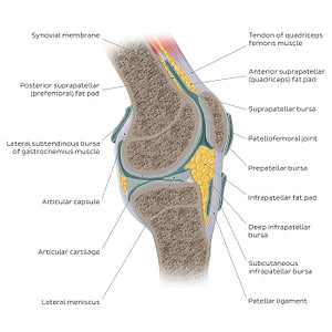
Sagittal view of the right knee joint, with all articulating surfaces clearly visible. The lateral and medial condyles of the femur articulate with the tibial plateau inferiorly forming the tibiofemoral joint. Anteriorly, the patellar surface of the femur articulates with the articular surface of patella forming the patellofemoral joint. This view of the knee joint is best for examining the structure of the articular capsule and its two parts, the outer fibrous layer and inner synovial membrane which encloses the articular cavity. The articular capsule forms several pouches called bursae, that cushion and reduce friction within the knee joint. Additional important structures are the menisci situated between the lateral and medial condyles of the femur and tibial plateau, providing congruency to these articulating surfaces. In the patellofemoral joint, one of the important supporting structures is the patellar ligament, that extends from the patella to the tibial tuberosity.
Normaler Preis
$7.56 USD
Normaler Preis
Verkaufspreis
$7.56 USD
Grundpreis
pro
Verfügbarkeit für Abholungen konnte nicht geladen werden

#E0AB37
#6D9499
#E0AB37
#385B57
#EED693 und #ACBAD5