
Paul Kim
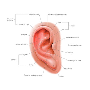
External ear: lateral view (English)
External ear: lateral view (English)
The auricle has several depressions and elevations that comprise its unique shape. The tragus is one of several cartilaginous flaps in the external ear and provides a lateral border to the distal end of the external acoustic meatus. The antitragus is located posteroinferior to the tragus, from which it is separated by the intertragic incisure. The helix forms the outer concave border of the ear and may present a small congenital protuberance called the auricular tubercle (of Darwin (not shown)). Internal to the helix in another raised cartilaginous structure called the antihelix which presents paired, fork-like crura at its superior extremity. It is separated from the helix by the scaphoid fossa. Finally, the inferiormost structure of the auricle is the soft, fibrofatty structure known as the lobule.
Normaler Preis
$7.56 USD
Normaler Preis
Verkaufspreis
$7.56 USD
Grundpreis
pro
Verfügbarkeit für Abholungen konnte nicht geladen werden

#E84C36
#B2695C
#6C1E12
#6C5856
#F59688 und #D1ABA2