
Paul Kim
Ankle joint: Medial view (English)
Ankle joint: Medial view (English)
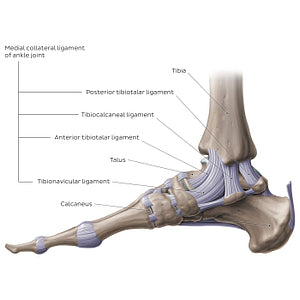
Considering its role in bearing the entire weight of the body, it is not surprising that the ankle joint has quite a few ligaments that stabilize it during movement. The ligaments are divided into two groups: the medial (tibial) and lateral (fibular) collateral ligaments. The medial collateral ligament, also known as the deltoid ligament, is a strong band that reinforces the medial aspect of the joint and prevents dislocations of the ankle joint. The ligament has a proximal attachment on the medial malleolus of the tibia, and fans out from there to insert onto the navicular bone, calcaneus, and talus. Consequently, the medial collateral ligament consists of 4 parts: the tibionavicular ligament, extending from the tibia to the navicular bone, the tibiocalcaneal ligament stretching from the tibia to the calcaneus, and anterior and posterior tibiotalar ligaments, extending from the tibia to the talus.
Normaler Preis
$7.56 USD
Normaler Preis
Verkaufspreis
$7.56 USD
Grundpreis
pro
Verfügbarkeit für Abholungen konnte nicht geladen werden

#8B796D
#624D3D und #C7B4A8